
Frequently Asked Questions
Glass Bottom Dishes
Mattek’s glass bottom dishes are available uncoated or coated with poly-d-lysine or collagen. All dishes are gamma irradiated to insure sterility.
A general procedure for their use follows.
- Maintain sterility: Open dishes in a sterile environment (e.g. laminar flow hood).
- Pre-equilibrate dishes: Incubate the dishes with culture medium. Pipet 2-3 ml of medium into the 35 mm dishes or 3-4 ml into the 50 mm dishes and incubate at 37° C for 15 minutes.
- Add cell suspension to microwell: Remove the culture medium by aspiration and plate cells onto the glass surface. Pipet 250 µl of the cell suspension (cells suspended in culture medium) into the 10 mm diameter microwells, 500 µl of cell suspension into the 14-mm microwells, or 1 ml of cell suspension into the 20-mm wells. Incubate the dishes for 1 hour at 37° C.
- Add additional medium: After 1 hour, gently fill the remainder of the dish with medium. Add 2-3 ml to the 35 mm dishes or 3-4 ml for the 50 mm dishes.
Note: After the initial one hour period to allow cells to attach to the glass surface, it is important to fill the dish to normal levels in order to minimize the effects of evaporation and to avoid inducing changes in osmolarity.
It is hard to predict which type of glass bottom dish will work best with your specific cell type. Uncoated (e.g. Part # P35G- or P50G-) or high adhesion (e.g. Part #: P35G-…..-HA) dishes work nicely for many transformed or cancerous cell lines. Poly-d-lysine coated dishes (Part #P35GC-…) work well for neuronal cultures and for many primary cells; other cells prefer a collagen coating (Part #P35GCol-…). Also, many researchers purchase our uncoated dishes and apply their own specialized coating. Conducting an online search for published methods utilizing Mattek glass bottom dishes and your cells may help determine your best choice (See FAQ #3 on how to search for published methods using Mattek glass bottom dishes and also references on our website.
Note: For almost all microscopy applications, the No. 1.5 coverslip thickness (e.g. Part #: P35G-1.5-… or P50G-1.5-…) is preferred.
We have collected and catalogued hundreds of technical papers that cite the use of Mattek Glass Bottom Culture Dishes. You can search our database of cataloged publications on our website here. Note: If the exact glass bottom dish part number is unspecified in a literature article, it is safe to assume that uncoated dishes were used (e.g. Part #’s: P35G-1.5-14-C, P50G-1.5-30-F, etc.)
To increase cell attachment and spreading, we recommend the high adhesion dishes (e.g. Part # P35G-1.5-14-C-HA).
- For some cells, optimal attachment and spreading will require an extracellular matrix coating. Our poly-d-lysine or collagen coating works well for the vast majority of cells, however, some customers find it necessary to coat the glass bottom dishes using their own specialized coating. These customers purchase the high adhesion (e.g. Part #: P35G-…..-HA) dishes and use the procedure along with their coating of choice.
- 1. Rinse the dish with phosphate buffered saline (PBS) and 1x with ultra-pure H2O;
- 2. Apply the coating of choice to the dishes. In many instances, allowing the coating solution to remain in contact with the glass for 18-24 hours at 37°C without letting the dish to go to dryness is beneficial. For neuronal cells, apply 0.5 mg/mL of poly-d-lysine overnight and do not allow the dish to dry
- 3. After coating is complete, add a similar volume of the medium in which you will plate your cells to pre-equilibrate the glass surface. Incubate the medium in glass bottom dishes for 15 min at 37°C. Remove the medium and then plate your cells.
To try a sample of non-coated glass bottom dishes, Request a free sample here.
The HA dishes utilize glass coverslips which have been specially cleaned and optimized for cell attachment and spreading. The high adhesion dishes (e.g. P35G-1.5-14-C-HA) are not coated with an extracellular matrix but many researchers find that the -HA dishes give better and more consistent attachment and growth of cells.
Yes, but for most applications, cells growing in the glass bottom dish can be viewed without removal of the coverslip using a variety of inverted and upright microscopes.
If necessary (e.g. for long-term storage purposes), the coverslip can be removed using the following procedure:
- Order Part #PDCF OS 30 (Fluid for removal of coverslips from glass bottom dishes)
- Invert the cover of the dish.
- Pipette 1.0 ml of fluid into the inverted cover.
- Place the bottom of the dish onto the cover. Make sure that the liquid in the inverted cover is touching the bottom of the coverslip.
- Allow the dish to sit in the fluid for 45 minutes at room temperature.
- Dry the bottom of the coverslip with an absorbent paper towel.
- Place the dish on a clean surface. Using forceps, press down on the inner edge of the coverslip to separate the coverslip from the dish.
Note: If the above procedure is followed, the PDCF OS 30 fluid will not contact the cells and will not disrupt cells on the coverslip or the staining thereof. Coverslips can be removed without breakage.
In order to approximate physiological conditions, the temperature of the medium contained within the glass bottom dishes can be controlled by using a microscope stage heater and an appropriate stage adapter.
For use with the P35G dishes (Corning 35 mm dishes): Culture dish heaters (part#: DH-35), microscope stage adapters (part#: SA-microscope type), heater controller (part#: TC324-B), and connector cable (part#: CC-28) are available from Warner Instrument Corporation. Information is available online at: https://www.warneronline.com/
The gridded coverslips allow one to refer to specific cells and follow them over time. For instance, individual cells can be microinjected, returned to the incubator, and observed at multiple time points since each cell can be identified with a unique alpha-numeric coordinate in the dish. Glass bottom dishes containing gridded Bellco Glass coverslips are available. Standard gridded dishes are designated as Part #’s: P35G-1.5-14-C-GRD and P50G-1.5-14-FGRD.
Grid size: The grid on Part #’s P35G-1.5-14-C-GRD and P50G-1.5-14-FGRD consists of 520 unique alphanumeric squares. Each square measures 600 microns x 600 microns. The line thickness is 20 microns.
Visualization of the grid: The grid should be readily observable using a 10X brightfield objective. After you locate the cell of interest, you can switch to a higher magnification or fluorescence objective. However, the grid will not be observable using higher power or fluorescence objectives.
I can’t see the grid: A confluent monolayer of cells will typically mask the grid making it difficult or impossible to visualize. If your cells are confluent, you can utilize Part # P35G-1.5-14-CGRD-D which places the grid on the outside of the dish where it is unaffected by the cells growing on the coverslip. To use this dish, find a cell of interest and then focus down to the bottom side of the coverslip to get its coordinates.
Correlative Light and Electron Microscopy (CLEM): The P35G-1.5-14-C-GRD glass bottom dishes work nicely with CLEM; however, the P35G-1.5-14-CGRD-D is not recommended for CLEM because the cells and the grid are in a different focal plane.
The glass bottom dishes can also be used to image tissue slices. The slices are adhered to the dishes using Corning Cell-Tak. A research paper utilizing Mattek’s glass bottom dishes to perform confocal microscopy on brain slices is available.
We do NOT recommend re-using the glass bottom dishes. The surface properties of the substrate on which cells are cultured have a profound effect on cell structure and function. Re-use of dishes will introduce uncontrolled variables into your experiments which may affect the phenomenon under study.
Mattek glass bottom dishes are meant for single-use experiments.
Mattek’s Glass Bottom Culture Dishes are standard size 35 and 50 mm disposable plastic petri dishes with glass coverslip bottoms, providing researchers with superior high resolution microscopic images of their in vitro cultures. These dishes are routinely used in confocal, polarized light, and fluorescence imaging techniques. These dishes are also ideal for live cell microscopy.
Mattek recently added Glass Bottom Multi-Well Plates to its glass bottom dish product offering.
Our dishes are known by many different names including glass bottom dishes, glass bottom culture dishes, glass bottom petri dishes, glass bottom microwell dishes, glass bottom sterile culture dishes, imaging dishes and coverslip bottom dishes. Regardless of what they are called, our 35 mm and 50 mm Glass Bottom Culture Dishes, and now our Glass Bottom Multi-well Plates, have become the de facto standard for high resolution microscopic imaging of in vitro cell cultures.
Mattek Glass Bottom Dish and Muti-well Plate part numbers include: P35G-0-10-C, P35GC-0-10-C, P35G-0-14-C, P35GC-0-14-C, P35G-0-20-C, P35GC-1.0-14-C, P35G-1.0-14-C, P35GC-1.5-10-C, P35G-1.5-10-C, P35GC-1.5-14-C, P35G-1.5-14-C, P35GCOL-0-14-C, P35G-1.5-20-C, P35GCOL-1.0-14-C, P35G-1.5-14-CGRD, P35GCOL-1.5-14-C, P50G-0-14-F, P50GC-0-14-F, P50G-1.5-14-F, P50GC-1.5-14-F, P50G-2-14-FGRD, P06G-0-20-F, P12G-0-14-F, P06G-1.0-20-F, P12G-1.0-14-F, P06G-1.5-20-F, P12G-1.5-14-F, P24G-0-13-F, P96G-1.5-5-F, P24G-1.0-13-F, P24G-1.5-13-F, P96GC-1.5-5-F.
Glass Bottom Dishes with gridded glass coverslips are also available. Part numbers include P35G-1.5-14-CGRD, P50G-1.5-14-FGRD.
Mattek Cell Culture Coverslip Kits
Mattek Cell Culture Coverslip Kits come ready to use – coverslips placed in 35 mm petri dishes and pre-sterilized. Standard glass coverslip kits are available in the CSGK/F, CSGK/C and CSGK/N Coverslip Kit configurations. These kits, the CSGK/F in particular, are widely used in amniocentesis testing.
Microscopy Types and Techniques
Yes. The glass bottom dishes are excellent for both confocal and fluorescent microscopy. Important glass properties are:
- Incident ultraviolet rays with wavelengths longer than 320 nm do not cause fluorescence.
- Mercury lines at 334 and 365 nm do not create auto-fluorescence. (Note: For mercury illumination, filter out the mercury lines with wavelengths shorter than 313 nm to obtain best possible results.)
- Refractive index (@ 20°C): ne 1.5255+/- 0.0015, nD 1.5230, ve 55
- Abbe number V = 55.
You will need to order BOTH a glass bottom dish and a glass cover. Order any P35G dish (e.g. Part #’s: P35G-1.5-14-C or P35G-1.5-20-C) along with a glass cover (Part #: P35GTOP-0-20-C). Both the glass bottom and glass cover are necessary so that the entire light path travels only through high optical quality glass. The glass covers can be re-used following re-sterilization by soaking them in 70% ethanol for 30 minutes. The covers CANNOT be autoclaved.
Yes. For high-resolution techniques such as GSDIM, dSTORM, PALM, TIRF, and for any objective with a numerical aperture > 0.7, tight control of the coverslip thickness is crucial. Therefore, use of Part # P35G-0.170-14-C dish which utilizes a high tolerance No. 1.5 coverslip (0.170 ± 0.005 mm) is necessary.
The 50 mm glass bottom dishes (part #’s beginning with P50G-) are useful for:
Microinjection: The larger diameter (50 mm) and the lower side wall (9 mm) allows easier access to cells in microinjection experiments.
Atmosphere maintenance: The 50 mm dish has a cover that snaps onto the dish bottom and thereby prevents loss of the 5% CO2 atmosphere while the dish is out of the incubator. This can be important for experiments in which dishes will be observed for extended periods.
Automate Scientific and Warner Instruments, Inc., make perfusion adapters which are compatible with Mattek’s 35 mm series of glass bottom dishes (Part #: P35G-xx-xx-C).
Dish Properties
Almost all objectives are optimized for a No. 1.5 coverslip thickness. Use of the No. 1.5 thickness gets increasingly important for higher numerical aperture coverslips (NA > 0.7). Use of other coverslip thicknesses can lead to optical distortion and loss of resolution. Therefore, for the vast majority of microscopy applications, the No. 1.5 coverslip thickness is optimal (e.g. Glass bottom dish Part #s: P35G-1.5-xx-C, P50G-1.5-xx-F, P60G-1.5-xx-F, etc.).
For super-high resolution microscopy techniques, we offer high tolerance No. 1.5 coverslips designated in part #s as -0.170 (e.g. Glass bottom dish Part #: P35G-0.170-14-C).
The actual thickness of the glass coverslips depends on the Coverslip No./Part #, as follows:
| Coverslip No./Part# | Thickness (mm) |
| 0 | 0.085-0.13 |
| 1.0 | 0.13-0.16 |
| 1.5 | 0.16-0.19 |
| 2.0 | 0.19-0.23 |
| 0.170* | 0.165-0.175 |
*Refers to Mattek designation in glass bottom dish Part #’s: P35G-0.170-14-C.
Although the specific identity of the adhesive is proprietary, the adhesive used is a non-toxic silicone that has been shown to be compatible with a broad variety of cells including primary neurons and many other difficult-to culture, fastidious cells.
Coated glass bottom dishes can be stored in the dark at room temperature for up to 1 year. Uncoated dishes can be stored for up to 5 years without any decline in cell growth properties.
The glass bottom dishes can be used over the temperature range -20°C to +45°C. The dishes will become disfigured at 55°C (131°F) and above. The glass bottom dishes CANNOT be autoclaved.
Mattek uses the highest quality, borosilicate German glass coverslips in its glass bottom dishes. The coverslip properties are as follows:
- Highest hydrolytic resistance (hydrolytic class 1).
- Excellent resistance to chemicals.
- Emission of alkali approximately 15 to 24 µg NA2O/g glass.
- Excellent properties for fluorescent microscopy.
- Incident ultraviolet rays with wavelengths longer than 320 nm do not cause fluorescence.
- Mercury lines at 334 and 365 nm do not create auto-fluorescence. (Note: For mercury illumination, filter out the mercury lines with wavelengths shorter than 313 nm to obtain best possible results.)
- Refractive index (@ 20°C): ne= 1.5255+/- 0.0015, nd= 1.5230
- Abbe number Ve = 55.
The depth of the micro-wells in the glass bottom dishes/ plates depends on the thickness of the dish/ plate bottom as follows:
- 35 mm dish (Part # P35G-…): 0.75-0.85 mm
- 50 mm dish (Part # P50G-…): 1.00-1.10 mm
- 60 mm dish (Part # P60G-…): 1.15-1.20 mm
- 100 mm dish (Par # P100G-…): 0.90-1.00 mm
- 6-well plate (Part # P06G-…): 1.45-1.55 mm
- 12-well plate (Part # P12G-…): 1.45-1.55 mm
- 24-well plate (Part # P24G-…): 1.10-1.20 mm
- 48-well plate (Part # P48G-…): 1.35-1.45 mm
- 96-well plate (Part # P96G-…): 1.05-1.25 mm
Note: For all dishes/ plates listed above, the sidewalls of each well contact the plastic bottom of the multi-well plate. To produce the glass bottom dish/ plate, a hole is cut into the bottom of each well to form a “micro-well.” The depth of the micro-well is equivalent to the thickness of the dish/ plate bottom, as listed above. In the glass bottom black 96-well plates and 384-well plates the sidewalls of each well directly contact the glass coverslip bottom (i.e. no micro-well is formed).
The body of the glass bottom dishes and multi-well plates is made from polystyrene. Therefore, they have limited compatibility with organic solvents. Please see the chemical compatibility table.
| Solvent | Chemical Compatibility |
| Acetone | Poor |
| Ammonium hydroxide (1N) | Fair |
| Ammonium hydroxide (25%) | Fair |
| Aniline | Good |
| Butanol | Good |
| Chloroform | Poor |
| Dimethylformamide | Poor |
| Dimethylsulfoxide(DMSO) | Poor |
| DMSO/H2O (20/80) | Good |
| Dioxane | Poor |
| Ethanol | Good |
| Hexane | Poor |
| Hydrochloric acid (25%) | Good |
| Hydrochloric acid (concentrated) | Fair |
| Methanol | Good |
| Methyl ethyl diketone | Poor |
| Methylene chloride | Poor |
| Nitric acid (25%) | Poor |
| Nitric acid (concentrated) | Poor |
| Sodium hydroxide | Good |
| Toluene | Poor |
| Xylene | Poor |
All glass bottom dishes are gamma irradiated at an FDA approved and certified vendor. We sterilize our dishes in bulk and typically >2000 separate cases are sterilized at the same time. Since sterility is an absolute requirement for all of our customers, the gamma dose that we use is excessive in order to ensure sterility. Following sterilization, dishes are subjected to quality control analysis to verify sterility: they are incubated in antibiotic- and anti-fungal-free medium for 7 days. In addition, each box has a gamma irradiation indicator that turns red upon exposure to gamma rays.
The P35G-0.170-14-C dishes utilize high tolerance No. 1.5 thickness coverslips (coverslip thickness = 0.170 +/- 0.005 mm) versus the standard P35G-1.5-14-C dishes (coverslip thickness = 0.175 +/ 0.015 mm).
The P35G-0.170-14-C dishes will improve picture quality (see Figure below) versus the P35G-1.5-14-C dishes for any high numerical aperture objective used in confocal, fluorescence, GSDIM, dSTORM, PALM total internal reflection (TIRF), and other high numerical aperture objectives. For example, quantitative measurements using P35G-0.170-14-C dishes gave z-resolution of +/- 9.5% while z-resolution in the P35G-1.5-14-C dishes gave z-resolution of +/- 17.3% (n=5).
| A | 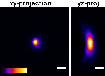 |
| B | 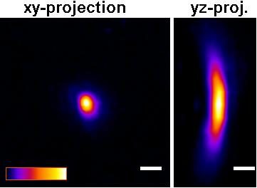 |
Figure: Improved Z-axis resolution – Effect of high tolerance glass coverslips (in P35G-0.170-14-C glass bottom dishes) on imaging of sub-resolution beads using:
A) P35G-0.170-14-C and B) P35G-1.5-14-C glass bottom dishes.
5 µl of FITC labeled 175nm PS-Speck sub-resolution beads were added to well of the glass bottom dishes and allowed to dry. After drying, 200 ul of water were added and the beads were imaged using a Zeiss LSM510 confocal microscope equipped with an Olympus UPLSAPO 60x (NA=1.2) water immersion objective.
Figures and measurements courtesy of Teemu Ihalainen, Ph. D., University of Jyvaskyla, Finland (2008).
Coatings
The poly-d-lysine used to coat the P35GC-x-xx-C and P50GC-x-xx-F glass bottom dishes is in the molecular weight range of 70,000-150,000 Daltons.
Both untreated glass and cells are negatively charged. Poly-lysine is applied to the glass surface to make it positively charged, thereby increasing electrostatic attraction between the glass surface and the cells and thus improving cell attachment. Poly-d-lysine is favored because the d-enantiomer is less prone to protease-mediated breakdown than the naturally-occurring l-enantiomer. Otherwise, Poly-d-Lysine and Poly-l-Lysine are equivalent. Mattek does not offer Poly-L-lysine coated dishes.
The collagen used to coat the P35GCol-x-xx-C or P50GCol-x-xx-F glass bottom dishes is type 1 rat tail collagen.
The coatings are monolayer coatings which do not affect the optical properties of the glass bottom dishes.
Special Orders
Glass bottom dishes with 7-mm diameter glass surfaces are standard products (e.g. Part # P35G-1.5-7-C or P35G-0-7-C). In addition, 50-mm, 60-mm- and 100-mm dishes come with a 30-mm diameter glass surface (e.g. Part #P50G-1.5-30-F, P60G-1.50-30-F, P100G-1.5-30-F) and are standard products; these dishes maximize the surface area for cell growth. When very expensive reagents need to be conserved, dishes with a 5-mm diameter glass surface (e.g. Part #: P35G-1.5-5-C) are also available on a special order basis.
Lead-time: Special order items typically can be produced within 2-3 weeks of receipt of a purchase order. However, bulk sterilization of the glass bottom dishes occurs only once every 6 weeks. Therefore, the lead-time for sterilized, special order products can vary between 3-8 weeks.
Note: Lead-times can be shortened if the customer will sterilizes the dishes using UV and/or 70% ethanol (e.g. 40 mins under UV light in a tissue culture hood and/or immersion if 70% ethanol for 30 minutes.
Special order charges: For special orders of ≥ 3 cases, there is no special order charge. However, for special orders of < 3 cases, a special order charge is assessed per case.
Return policy: Special orders items cannot be returned.
A: The main advantage of the multi-well glass bottom plates is that you can grow 6, 12, 24, 48 or 96 cultures under identical conditions in the same culture plate. The multi-well plates are ideal for high throughput, high/high content screening applications.
B: Analysis using the multi-well plates is streamlined because only one (1) plate (versus multiple petri dishes) needs to be handled.
C: For a number of applications, treatment of the cultures (e.g., irradiation) is simplified using the multi-well plates.
D: Smaller wells in the glass bottom multi-well plates are useful for application of precious reagents in smaller volumes.
Yes. In order to minimize back-scattered light and background fluorescence, we offer 96-well glass bottom plates (part # PBK96G-1.5-5-F) that are black. All properties of the PBK96G-1.5-5-F plates are identical to the standard, clear-wall 96-well glass bottoms plates (Part #: P96G-1.5-5-F) except the sidewalls of each well in the PBK96G-1.5-5-F plates are opaque (black). Note: Back-scattered light and background fluorescence is not a problem in the larger well 6-well, 12-well, 24-well, and 48-well glass bottom plates.
Mattek Chambered Cell Culture Slides
| Product | Dimensions of wells (cm) | Growth area (cm2) | Medium volume/ well (mL) |
| CCS-2 | 2.17 x 2.09 | 4.55 | 1.2 – 2.5 |
| CCS-4 | 2.15 x 0.99 | 2.13 | 0.5 – 1.3 |
| CCS-8 | 1.00 x 0.98 | 0.98 | 0.2 – 0.6 |
Yes, cells grown on the CCS can be observed using an inverted scope. Stain your cells and remove the plastic upper chamber. Use mounting medium and coverslip the samples on the slide. Mattek coverslips, Part #: PCS-1.5-5024 (No. 1.5 thickness 50 mm x 24 mm), are specially designed for coverslipping samples cultured on the CCS.
The dimensions of microscope slide are: Length: 75.0 mm, Width: 25.0 mm, and Height: 1.0 mm.
There is a silicone gasket beneath the upper chamber and the microscope slide. The gasket typically releases from the slide when the chamber is removed. However, if the gasket remains on the microscope slide, it can easily be peeled off the microscope slide using fine point forceps.
Although we do not produce Cell Culture Slides with more than 8 wells Mattek Glass Bottom plates are available with 12, 24, 48, 96, and 384 wells.
No. The CCS are not coated but the high purity soda lime glass used will support the growth of many cell types. For cells that are more fastidious, an ECM coating can be added by the end user. Sterile ECM coatings should be applied fresh for optimal cell attachment and growth. After applying the ECM coating under sterile conditions, rinse 2X with phosphate buffered saline and 1X with the culture medium to be used for cell growth, prior to seeding your cells. Do not let the ECM coating go to dryness.
Glass Coverslips
Mattek’s high quality German Glass Coverslips provide the optimal thickness to use for high resolution microscopy techniques. In particular, when using a high Numerical Aperture (N.A.) Mattek Glass Coverslips are ideal for:
- Confocal Microscopy Confocal Laser Scanning Microscopy (CLSM)
- Fluorescence Microscopy
- Green Fluorescence Protein (GFP)
- Fluorescence Resonance Energy Transfer (FRET)
- Immunofluoresence Microscopy
- Phase Contrast Microscopy
- Polarized Light Microscopy
- Differential Interference Contrast (DIC)
- Multi Photon Laser Scanning Microscopy (MPLSM)
- High Resolution Image Analysis
- Infrared Imaging
- Calcium Imaging
… and Mattek Glass Coverslips (in particular, part #: PCS-170-1818) have been used by customers for super-high resolution techniques such as:
- Ground State Depletion with Imaging Microscopy (GSDIM)
- direct stochastic optical reconstruction microscopy (dSTORM)
- Photoactivated localization microscopy (PALM)
- Total internal reflection (TIRF)
Almost all objectives are optimized for a No. 1.5 coverslip thickness. Use of the No. 1.5 thickness gets increasingly important for higher numerical aperture coverslips (NA > 0.7). Use of other coverslip thicknesses can lead to optical distortion and loss of resolution. Therefore, for the vast majority of microscopy applications, the No. 1.5 coverslip thickness is optimal (Coverslip Part # PCS-1.5-xx.). The actual thickness of the glass coverslips depends on the Coverslip No./Part #, as follows:
| Coverslip No. | Part# | Glass Thickness (mm) |
| 0 | PCS-0-xx | 0.085-0.13 |
| 1 | PCS-1.0-xx | 0.13-0.16 |
| 1.5 | PCS-1.5-xx | 0.16-0.19 |
| 2.0 | PCS-2.0-xx | 0.19-0.23 |
| 1.5 | PCS-170-xx | 0.165-0.175 |
Yes! High tolerance Mattek Glass Coverslips (part # PCS-170-1818) are optimized for super-high resolution techniques such as GSDIM, dSTORM, PALM total internal reflection (TIRF), and confocal microscopy using high numerical aperture objectives.
Many cells will grow directly on Mattek uncoated coverslips. However, more difficult to grow cell types (e.g. neurons) will require coating with an extracellular matrix coating. Please contact Customer Service for assistance.
The gridded coverslips allow one to refer to specific cells and follow them over time. For instance, individual cells can be microinjected, returned to the incubator, and observed at multiple time points since each cell can be identified with a unique alpha-numeric coordinate on the coverslip. Mattek’s gridded Glass Coverslips are available under part# PCS-1.5-1818-GRD.
Grid size: The grid consists of 520 unique alphanumeric squares. Each square measures 600 μm x 600 μm. The line thickness is 20 μm.
Visualization of the grid: The grid should be readily visible using a 10X brightfield objective. After you locate the cell of interest, switch to a higher magnification or fluorescence objective. However, the grid will not be visible using higher magnification or fluorescence objectives.
I can’t see the grid: A confluent monolayer of cells will typically mask the grid making it difficult or impossible to visualize. If you plan to visualize individual cells in a confluent monolayer, plate the cells on the side of the coverslip opposite the grid. To use this method, simply find a cell of interest and then focus down to underside of the coverslip to get the cell’s coordinates.
- Highest hydrolytic resistance (hydrolytic class 1).
- Excellent resistance to chemicals.
- Emission of alkali approximately 15 to 24 µg Na2O/g glass.
- Excellent properties for fluorescent microscopy.
- Incident ultraviolet rays with wavelengths longer than 320 nm do not cause fluorescence.
- Mercury lines at 334 and 365 nm do not create auto-fluorescence. (Note: For mercury illumination, filter out the mercury lines with wavelengths shorter than 313 nm to obtain best possible results.)
- Refractive index (@ 20°C): nD ** = 1.5230 tolerance ± 0.0015.
- Abbe number V = 55.
No. Mattek glass coverslips are shipped non-sterile and should be autoclaved prior to use. After sterilization, place the coverslips into a sterile petri dish or multi-well plate, and plate your cells.
Multiwell Plates
Plate Utilization
Mattek’s glass bottom multi-well plates are routinely utilized for fluorescence/immunofluorescence microscopy, confocal, live cell imaging, microinjection, videomicroscopy, phase contrast and time lapse photomicroscopy. Our glass bottom multi-well plates can be used for almost all of the same applications for which the standard glass bottom dishes can be used.
- The main advantage of the multi-well glass bottom plates is that you can grow 6, 12, 24, 48 or 96 cultures under identical conditions in the same culture plate. The multi-well plates are ideal for high throughput, high/high content screening applications.
- Analysis using the multi-well plates is streamlined because only one (1) plate (versus multiple petri dishes) needs to be handled.
- For a number of applications, treatment of the cultures (e.g., irradiation) is simplified using the multi-well plates.
- Smaller wells in the glass bottom multi-well plates are useful for application of precious reagents in smaller volumes.
Pharmaceutical Companies (due to high throughput testing), Academic/Research Institutions, and Medical Research Institutions.
Feedback has been very positive. A number of customers have purchased multi-well plates without first trying a sample based on their previous experience with the standard glass bottom dishes.
For new users, a sample plate can be requested on our free sample page.
The 6-well glass bottom plate can be used with upright or inverted microscopes. However, the 12-well, 24-well, 48-well and 96-well plates are compatible only with inverted microscopes.
Yes. However, for most applications, cells growing in the glass bottom multi-well plates can be viewed without removal of the coverslips. If necessary (e.g. for long term storage purposes), the coverslips can be removed using the following procedure:
- Order part # PDCF OS 30 (Fluid for removal of coverslips from glass bottom plates, available from Mattek).
- Place the cover of the multi-well plate on a clean surface top side up (Do not invert the cover).
- Pipette 3.0 ml of PDCF OS 30 fluid onto the top side of the cover.
- Place the bottom of the multi-well plate on top of the cover. The coverslip removal liquid on the cover should be touching the coverslips attached to the bottom of the multi-well plate.
- Allow the plate to sit in the fluid for 45 min at room temperature.
- Blot the bottom of the multi-well plate using an absorbent paper towel.
- Place the multi-well plate on a clean surface. Using forceps, press down on the edge of the coverslips to separate the coverslips from the plate.
Note: If the above procedure is followed, the PDCF OS 30 fluid will not contact the cells and will not disrupt cells on the coverslip or the staining thereof. Coverslips can be removed without breakage.
For long term storage of samples, Mattek chambered cell culture slides (available in 2-well, 4-well, and 8-well slides) are an excellent option.
You will need to order BOTH a glass bottom multi-well plate and a multi-well glass cover. Order any P06G plate (e.g. part #: P06G-1.5-20-F), P12G plate (e.g. part #: P12G-1.5-14-F), or P24G plate (e.g. part #: P24G-1.5-13-F) along with a glass cover (e.g. part# P24GTOP-1.5-F). The glass covers can be re-used following re-sterilization of the covers by soaking them in 70% ethanol for 30 minutes.
Mattek glass bottom multi-well plates are intended for single-use experiments.
We do NOT recommend re-using the glass bottom multi-well plates. The surface properties of the substrate on which cells are cultured have a profound effect on cell structure and function. Reuse of plates will introduce uncontrolled variables into your experiments which may affect experimental results.
Plate Properties
The depth of the micro-wells and the height of the sidewall depends on the type of multi-well plate:
- 6-well plate: Microwell depth = 1.45-1.55 mm; Well depth = 17 mm
- 12-well plate: Microwell depth = 1.45-1.55 mm; Well depth = 17 mm
- 24-well plate: Microwell depth = 1.10-1.20 mm; Well depth = 17 mm
- 48-well plate: Microwell depth = 1.05-1.20 mm; Well depth = 18 mm
- 96-well plate: Microwell depth = 1.05-1.25 mm; Well depth = 10 mm
- 6-well plate: 10, 14, or 20 mm
- 12-well plate: 10 or 14 mm
- 24-well plate: 10 or 13 mm
- 48-well plate: 6 mm
- 96-well plate: 5 mm
- 6-well plate: 3-4 ml / well
- 12-well plate: 1-2 ml / well
- 24-well plate: 0.5-1.0 ml / well
- 48-well plate: 0.25-0.35 ml / well
- 96-well plate: 0.1-0.2 ml / well
Although the specific identity of the adhesive is proprietary, the adhesive used is a non-toxic silicone that is compatible with a broad variety of cells including primary neurons and many other difficult-to culture, fastidious cells.
The body of the glass bottom dishes and multi-well plates is made from polystyrene. Therefore, they have limited compatibility with organic solvents. Please see the chemical compatibility table.
| Solvent | Chemical Compatibility |
| Acetone | Poor |
| Ammonium hydroxide (1N) | Fair |
| Ammonium hydroxide (25%) | Fair |
| Aniline | Good |
| Butanol | Good |
| Chloroform | Poor |
| Dimethylformamide | Poor |
| Dimethylsulfoxide(DMSO) | Poor |
| DMSO/H2O (20/80) | Good |
| Dioxane | Poor |
| Ethanol | Good |
| Hexane | Poor |
| Hydrochloric acid (25%) | Good |
| Hydrochloric acid (concentrated) | Fair |
| Methanol | Good |
| Methyl ethyl diketone | Poor |
| Methylene chloride | Poor |
| Nitric acid (25%) | Poor |
| Nitric acid (concentrated) | Poor |
| Sodium hydroxide | Good |
| Toluene | Poor |
| Xylene | Poor |
All glass bottom multi-well plates are gamma irradiated at an FDA approved and certified vendor. We sterilize our dishes in bulk – typically >5000 separate cases are sterilized at the same time. Since sterility is an absolute requirement for all of our customers, the gamma dose that we use is excessive in order to ensure sterility.
Following sterilization, dishes are subject to our quality control analysis to verify sterility: they are incubated in antibiotic- and anti-fungal-free medium for 7 days. In addition, each box has a gamma irradiation indicator which turns red upon exposure to gamma rays.
Yes. In order to minimize back-scattered light and background fluorescence, we offer 96-well glass bottom plates (part # PBK96G-1.5-5-F) that are black. All properties of the PBK96G-1.5-5-F plates are identical to the standard, clear-wall 96-well glass bottoms plates (part #: P96G-1.5-5.F) except the sidewalls of each well in the PBK96G-1.5-5-F plates are opaque (black).
Note: Back-scattered light and background fluorescence is not a problem in the larger well plates (i.e. 6-well, 12-well, 24-well, and 48-well glass bottom plates).
Plate Coatings
Currently, only poly-d-lysine coated 96-well plates are available as standard products (Part #’s: P96GC-1.5-5-F and P96GC-0-5-F).
PermaCell Cell Culture Inserts
The PermaCell inserts and 96-well insert plates are designed for 3-dimensional organotypic tissue culture. Cells are seeded into the insert onto the apical side of the microporous membrane. Cultures can then be fed submerged (medium added to basal and apical sides of the membrane or at the air-liquid interface (ALI) where medium in only added to the basal compartment (i.e. the apical side of the culture is exposed to the atmosphere in the incubator). For many cell types, culture at the ALI improves cellular differentiation and induces formation of in-vivo like, 3-dimensional tissue properties.
The Hanging Top Plate allows you to use a higher volume of medium in the basal compartment, reducing the frequency of media exchanges. Inserts can be hung in a 12-well plate (Part #: CCI12-HANGTOP) or a 24-well plate (Part #: CCI24-HANGTOP). If using the CCI12-HANGTOP plate, fill each well with 5.0 mL of medium. If using the CCI24-HANGTOP plate, fill each well with 2.5 mL of medium. For the 96-well insert plate, use 0.3 mL per well (no Hanging Top Plate is necessary).
PermaCell inserts are available with different membrane material options:
a) polytetrafluorethylene (PTFE, Part # CCI24-PTFE-0.4)
b) polyethylene terephthalate (PET, Part # CCI24-PET-0.4)
c) polycarbonate (PC, Part # CCI24-PC-0.4).
All membrane options have high pore density (pore size 0.4 µm, 1 x 10⁸ pores/cm2). Different cell types may behave differently on each of these membranes. If you are not sure which type of insert to use, a trial pack which contains 4 inserts each of the PTFE, PET, and PC inserts is available (part #: CCI24-trial). Note: One potential advantage of the PTFE inserts over the PET and the PC inserts is that the PTFE membrane is microscopically transparent when wet. Thus, cells growing on the PTFE membrane can be observed using an inverted microscope. For higher throughput testing, use the PermaCell 96-well insert plates (Part # CCI96-PET-0.4 or CCI96-PET-CL-0.4).
The PermaCell inserts are tissue culture treated and will support growth and differentiation of many cell types without an ECM coating. However, depending on your cell type and endpoints/ tissue properties of interest, an ECM coating may be beneficial. A coating volume of 150-250 µL/cm2 is recommended for most applications.
A non-destructive convenient way to monitor the formation of a confluent cell layer or tissue on the inserts is to measure the transepithelial electrical resistance (TEER). We recommend the EndOhm-12 chamber from World Precision Instruments for use with CCI24 PermaCell inserts. For the PermaCell 96-well insert plate, we offer a specially designed TEER probe (Part #: CCI96-TEER).
After formation of a confluent cell/tissue layer, all PermaCell inserts and insert plates are appropriate for permeability experiments. The donor solution can be added directly to the apical tissue surface and permeation into the basal solution can be monitored over time. For chemotaxis experiments looking at passage of cells through the tissue into the basal compartment, inserts with the 8µm pore polycarbonate membrane (part # CCI24-PC-8) should be ordered. This membrane will allow cells to migrate through the tissue and the underlying membrane into the basal compartment.
The polytetrafluorethylene (PTFE, Part # CCI24-PTFE-0.4) inserts and the clear polyethylene terephthalate (PET, Part # CCI24-PET-CL-0.4) insert plates are transparent in aqueous media. Cells cultured on the inserts can be viewed directly using an inverted microscope with 10X or 20X objectives.
About Mattek
Mattek Corporation was founded in 1985 by two MIT chemical engineering professors. In 1991, the company leveraged its core polymer surface modification technology into the emerging tissue engineering market. In 1993, EpiDerm, Mattek’s first in vitro human tissue equivalent, was introduced and has been in continuous production from 1993 to the present.
Mattek produces a variety of normal (non-transformed), human cell-derived, 3-dimensional, organotypic in vitro tissue equivalents. This Web site, Mattek.com, is devoted to providing detailed information about our in vitro human tissue equivalents. They are mitotically and metabolically active, closely mimic their in vivo counterparts, both structurally and biochemically, and do so with guaranteed reproducibility.
Mattek also produces a line of Glass Bottom Culture Dishes. These dishes are used in confocal, polarized light, and fluorescence microscopy techniques, and are ideal for live cell microscopy applications.
3D Tissue Models
Mattek produces 12 lab-grown human tissue models, all of which are derived from human epithelial cells (click on the tissue name for detailed information):
- EpiDerm, our human epidermal tissue equivalent, consists of normal, human cell-derived epidermal keratinocytes that have been cultured to form a multilayered, highly differentiated model of the human epidermis. EpiDerm, also known generically as Reconstructed Human EpiDermis (RhE), has been in continuous production for over 15 years. There is also an “under-developed” version, EpiDerm-201
- EpiDermFT (EpiDerm Full Thickness), a dermal / epidermal human skin equivalent with a well-defined, fully functional basement membrane.
- MelanoDerm is a human skin equivalent composed of keratinocytes and melanocytes that have been cultured to form a multilayered, highly differentiated model of the human epidermis.
- Melanoma is a full thickness melanoma skin model composed of human malignant melanoma cells, normal, human-derived epidermal keratinocytes and normal, human-derived dermal fibroblasts that have been cultured to form a multilayered, highly differentiated epidermis with melanoma cells at various stages of CM malignancy.
- EpiOcular consists of normal, human-derived epidermal keratinocytes which have been cultured in serum free medium to form a stratified, squamous epithelium, similar to in vivo human corneal epithelium
- EpiCorneal consists of normal human corneal epithelial cells cultured to form a stratified, squamous epithelium which closely parallels normal human corneal tissue.
- EpiAirway consists of normal, human cell-derived tracheal/bronchial epithelial cells that have been cultured to form a pseudo-stratified, highly differentiated model that closely resembles the epithelial tissue of the respiratory tract.
- EpiAlveolar is produced from primary human alveolar epithelial cells, pulmonary endothelial cells and fibroblasts.
- EpiOral, our human buccal (inner cheek) equivalent,
- EpiGingival, our human gingival (gum) tissue equivalent.
- EpiVaginal, derived from human ectocervico-vaginal (ECV) epithelial cells.
- EpiIntestinal, incorporates enterocytes, paneth cell, M cells, tuft cells and intestinal stem cells into a highly differentiated, polarized epithelium
Typical configurations include:
- 9 mm diameter individual tissues, each tissue produced in a single well tissue culture plate insert (shipped in either 12 or 24 tissue kits with media)
- 22 mm diameter individual tissues, each tissue produced in a single well tissue culture plate insert (shipped in 6 tissue kits with media)
Tissues are also available in 24-well and 96-well high throughput plates. Special configurations are also available for specific tissue types (ex. EpiAirway tissues produced in “snapwell” culture inserts for use in Using Diffusion Chambers).
Yes, Mattek also produces Human Dendritic (Langerhans) Cells. Mattek has developed a new method for generating Human Dendritic Cells from CD34+ progenitor cells (hematopoietic stem cells) harvested from umbilical cord blood. Applications include allergenicity, antigen binding and presentation, viral infection, immuno-therapeutic and transfection studies.
Yes. EpiVaginal with Langerhans Cells (“VLC” products) is a released product. Several other Mattek in vitro human tissue equivalents that incorporate Dendritic Cells are in “beta” phase. Please contact Mattek for more detailed information.
There are many applications for each model. For example, our EpiDerm in vitro skin equivalent is used to determine and/or study the following: dermal corrosion, skin irritation (cutaneous toxicity), dermal phototoxicity, percutaneous absorption (drug permeability, transdermal drug delivery), inflammation, gene analysis, antioxidants, metabolism, apoptosis, antimicrobial peptides, and angiogenesis.
A very useful method to determine if Mattek In Vitro Products have been used in applications similar to your application is to search our list of Technical References.
Pharmacology/Toxicology pre-clinical applications for our in vitro human tissue equivalents include transdermal, transbuccal, transmucosal drug delivery; biocompatibility, toxicity studies; HIV, microbicide research; bioequivalence studies; lead optimization, etc.
An example of a pharm/tox application of Mattek’s in vitro human tissue equivalents is as follows: after a new drug library has been processed thru a biochemical-based (enzyme) primary screening and a cell-based secondary screening, but PRIOR to performing pre-clinical animal studies, the remaining drug candidates are passed through a tertiary in vitro human tissue-based screening to determine optimum permeability and/or minimal toxicity characteristics. Those drug candidates that pass this tertiary screening then move onto the animal-based study, but that study will now require fewer animals, and can therefore be structured as a small confirmatory study to meet FDA (or other regulatory agency) requirements. Also, by performing the human tissue-based tertiary study prior to commencement of human clinical studies, the potential for cross-species extrapolation errors based on animal study results has also been significantly reduced.
Prior to purchasing our in vitro human tissue equivalents, you can purchase purified total RNA (control and treated) and/or Protein Lysate (control) from each of our human tissue equivalents to confirm the expression level of specific gene(s) or the presence/absence of specific protein(s).
Mattek’s in vitro human tissue equivalents are very reasonably priced, especially considering the costly materials used to produce these tissues, and the amount of time (2-3 weeks) and labor (numerous technician “touches” per lot) required to grow a given tissue. The cost of these tissues is considerably less than the cost of the end user trying to produce these tissue equivalents reproducibly from initial harvesting of seed cells. Printed price lists are available upon request from Mattek—to receive one, just complete our online Information Request Form.
No, none of Mattek’s in vitro human tissue equivalents are approved for human use.
With one exception (Japan), Mattek does not have dealers or distributors for its in vitro products. The nature of the product is such that it is best if end-users deal directly with Mattek.
Typically, because of the nature of the production process, Mattek does not provide free samples of our in vitro human tissue equivalents. Mattek will provide proof of concept to qualified potential Customers. Please contact Mattek for details.
Tissues are typically shipped at 4°C on medium-supplemented agarose gel.
Shipment day: Every Monday.
Shipment Delivery: Tuesday morning via FedEx priority service (US). Outside US: Tuesday-Thursday depending on location.
Yes, Mattek has many international Customers, particularly in the European Union. Each country has a unique set of shipping requirements. Please contact Mattek for more information about shipping requirements for a particular country.
Mattek can do a proof of concept for your organization–please contact Mattek for details. Once we have a conversation about your intended application, we can also send you a list of Mattek-approved Contract Testing Labs that can perform assays using Mattek in vitro human tissue equivalents.
Q84: Are there detailed protocols that I can follow to use Mattek in vitro human tissue equivalents?
Yes, Mattek has detailed protocols for all of the major applications for each of our in vitro human tissue equivalents. Please contact Mattek with your specific application and we will provide the appropriate protocol for your review.
Normal Cells: Non-transformed cells. Mattek does not use immortalized cell lines. By definition, an immortalized cell line has been transformed, and therefore possesses characteristics that are not desirable in a true in vitro human tissue equivalent.
Human Cell-Derived: All Mattek tissue equivalents are derived from human cells. This procedure eliminates the cross-species extrapolation concerns that accompany all work done using non-human cells.
Organotypic: Mattek’s production process produces differentiated, multi-layer, 3-D human tissue equivalents that closely resemble those found in vivo. Mattek advances in tissue engineering have made it feasible to use in vitro human tissue equivalents to explore many of the scientific questions that could only be pursued in vivo previously.
Reproducibility. No other feature is more important for the successful use of in vitro human tissue equivalents than reproducibility. Why? Without proven reproducibly, an organization can not use this technology in applications that are designed to run over an extended period of time. For example, if your organization plans to use in vitro human tissue equivalents to build a database of dermal irritation information about its product formulations, that database will remain useful over many years only if it is based on data from assays that use highly reproducible in vitro human tissue equivalents.
For this reason, Mattek has spent many years developing the most reproducible in vitro human tissue equivalents available. For example, Mattek has over 15 YEARS of reproducibility data for the EpiDerm human skin equivalent.
Reproducibility is so important to the successful use of in vitro human tissue equivalents that Mattek GUARANTEES the reproducibility of ALL of its in vitro tissues.
Complete the Mattek Information Request Form including your detailed application information. We will review your application information and recommend the appropriate in vitro human tissue equivalent and protocol for that application. The more detailed you are in the description of how you intend to use our products in your work, the more specific we can be in our to response to your inquiry.
“TR” indicates a Mattek Technical Reference. Technical References are technical posters/papers that scientific staff members at Mattek and/or at organizations using Mattek in vitro products have presented at major technical conferences and meetings.
There is a searchable database of TR citations/summaries on the Mattek Web site. There are over 700 TR’s (and growing!) listed in the database. Click here to go to the Mattek TR page.
As mentioned above, there is a searchable database of Technical Reference (TR) citations/summaries on the Mattek Web site. There are over 700 TR’s listed in the database. Click on this link to go to the Mattek TR page.
ICCVAM (USA) and ECVAM (Europe) are the organizations most directly responsible for the effort to introduce and validate non-animal alternative toxicological test methods in order to eliminate/reduce the number of animals used in these studies.
From the ICCVAM Web site: “The Interagency Coordinating Committee on the Validation of Alternative Methods (ICCVAM) was established in 1997 by the Director of the National Institute of Environmental Health Sciences (NIEHS) to implement NIEHS directives in Public Law (P.L.) 103-43. This law directed NIEHS to develop and validate new test methods, and to establish criteria and processes for the validation and regulatory acceptance of toxicological testing methods. P.L. 106-545, the ICCVAM Authorization Act of 2000, established ICCVAM as a permanent committee. The Committee is composed of representatives from 15 Federal regulatory and research agencies; these agencies generate, use, or provide information from toxicity test methods for risk assessment purposes. The Committee coordinates cross-agency issues relating to development, validation, acceptance, and national/international harmonization of toxicological test methods.”
From the ECVAM Web site: “ECVAM (European Centre for the Validation of Alternative Methods) was created by a Communication from the Commission to the Council and the Parliament in October 1991, pointing to a requirement in Directive 86/609/EEC on the protection of animals used for experimental and other scientific purposes, which requires that the Commission and the Member States should actively support the development, validation and acceptance of methods which could reduce, refine or replace the use of laboratory animals. ECVAM was established in 1992 as a unit of the Environment Institute, part of the Joint Research Centre, and was transferred, at that time, to the newly formed Institute for Health and Consumer Protection in Ispra, Italy in 1998.”
From: The Principles and Procedures of Validation, Chapter 2 (ATLA 30, Supplement 1, 13.19, 2002): “The validation of an alternative method is the process by which the relevance and reliability of the method are established for a particular purpose. In the context of a replacement test method, relevance refers to the scientific basis of the test system, and to the predictive capacity of an associated prediction model (PM), whereas reliability refers to the reproducibility of test results, both within and between laboratories, and over time.”
The entire validation process for an alternative method can take 8-10 years. The scientific portion of the process (formal pre-validation and validation studies) usually takes 4-6 years.
Mattek has TWO alternative methods formally validated – EpiDerm for skin irritation testing and for dermal corrosion testing. Several others have completed major portions of the process – EpiOcular for ocular irritation (an alternative to the Draize Rabbit Eye Test) that is being sponsored by Colgate-Palmolive, and EpiDerm for percutaneous absorption testing.
The MTT Assay is a very accurate end point (method) used to measure the cell viability of in vitro human tissue equivalents. The assay is a colorimetric assay that measures the reduction of a tetrazolium component (MTT) into an insoluble formazan product by the mitochondria of viable cells. After incubation of the cells with the MTT reagent for several hours, a solution is added to lyse the cells and solubilize the colored crystals. Samples are read using an ELISA plate reader at a wavelength of 570 nm. The amount of color produced is directly proportional to the number of viable cells.
Yes, Mattek’s EpiDerm in vitro human tissue equivalent has been used successfully in several such studies. Click on the following link to review information related to this topic: Transdermal Drug Delivery
Yes, Mattek’s EpiAirway tracheal/bronchial in vitro human tissue equivalent has been used successfully in several intranasal and inhaled drug delivery studies. Click on the following links to review information related to Drug Delivery.
Organ-on-a-Chip Culture Platform
No additional equipment is needed. AIM chips and plates are standalone consumables akin to standard tissue culture plasticware. If you currently perform cell culture experiments in your lab, all you’ll need to procure is a polymerizable hydrogel of your choice to extend your studies into 3D.
To date, there are more than 80 publications by our customers using AIM Biotech devices. Over 60 publications have also been made by the scientific founders of AIM Biotech and their collaborators using non-commercial versions of the technology. Publications by AIM Biotech customers using the commercial chips have been accepted into prestigious journals like Nature Medicine, Cell, Cancer Cell etc. The full list of publications is available here. Their studies encompass immuno-oncology (immune checkpoint & T cell therapy), blood-brain barrier, angiogenesis, transendothelial migration, cancer aggregate dispersion, cancer cell migration, extravasation, intravasation, neurite guidance, stem cell differentiation, etc. AIM Biotech idenTx Chips/Plates are open platforms for you to test new ideas and to develop interesting applications. The idenTx family of chips & plates feature 1 gel channel flanked by 2 media channels. In the near future, AIM will be releasing chips with different channel configurations that will enable new experimental designs.
Any polymerizable hydrogel that suits your application can be used, e.g. various types of collagen, fibrin, etc. The hydrogels may be used on their own or in combination with other components like laminin, Matrigel, etc. However, do note that hydrogel quality may vary from manufacturer to manufacturer. There may also be batch-to-batch variations.
The optimal cell density depends on your specific application and the design of your experiment. For example, an idenTx 3 chip (with 3 microfluidic sites per chip) will need approximately 30,000 endothelial cells per site to generate a confluent endothelial monolayer overnight. By contrast, you will only need 3000 cells per site if you plan to seed the cells in the hydrogel to study cell migration. AIM chips are flexible platforms that can be adapted to various cell seeding densities; you may have to optimize your cells seeding strategies for your specific applications. Please visit the Applications-Specific Protocols section of this website for more information on specific applications.
Unlike standard 2D cell culture flasks, microfluidic devices use very small quantities of culture media. As such, you should change media daily to replenish the consumed media. We recommend that users attach a syringe pump to maintain a stable gradient or interstitial flow.
AIM chips can be connected to devices with luer interfaces through AIM Luer Connectors (LUC-1). The connectors attach directly into media channel ports to form a female luer lock interface, enabling users to connect male luer components such as:
• syringe barrels – to form media reservoirs, or to establish pressure gradients for interstitial flow across the gel channel
• syringe pumps via tubing and 3rd party male luer tubing connectors – to run dynamic cell culture experiments, to enable media exchange or to set up interstitial flow or chemical gradients across the gel channel
Please use the included Inlet Seals to seal off the gel inlets. This will stop cell culture media from flowing through the porous gel and out through the gel inlets.
AIM Biotech idenTx Chips/Plates can be viewed under bright field, phase contrast, epi-fluorescence, 2-photon and confocal microscopy. The chips are also compatible with oil/water immersion techniques for high magnification imaging. However, do note that signals may become weaker when a high numerical aperture (NA) lens is used to image deeper regions of the chips due to scattering & spherical aberration.
You may pool the media in the media channels from multiple sites and detect analytes (typically proteins of interest) present in the media through the enzyme-linked immunosorbent assay (ELISA) or other assays. However, do note that the local concentration of a substance in the channel is usually higher than the concentration in the media that you collect from the ports.
Cells in the media channel can also be collected by trypsinization. Hydrogels can also be dissolved with the appropriate enzymes (e.g. trypsin for fibrin gels, collagenase for collagen gels) to release cells from within the gel. You can perform polymerase chain reaction (PCR) or other assays that utilize collected cells as an input. Do take into account the number of cells that can be collected from each channel for your experimental setup. You may need to adjust your collection strategy for assays that require more cells.
No, the AIM Biotech idenTx Chips/Plates are not designed to be reused. The sterility and functionality of the chips cannot be guaranteed if the chips are used again.
Yes, you can. However, you have to make sure the chips are elevated so that gas exchange can take place through the laminated underside. The microtiter plate holder is designed to ensure the air circulation is not obstructed on the laminated underside. The holder also helps to reduce the evaporation loss of media from the chips.