
Respiratory Infection
Use EpiAirway to study respiratory pathogen attachment, replication, innate immune responses and anti-viral/anti-bacterial drug development.
METHODS
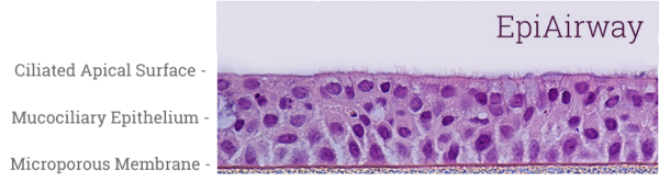
EpiAirway tissues (Figure 1) equilibrate overnight under standard culture conditions (37°C, 5% CO2) with EpiAirway Assay Medium* (AIR-100-ASY). After 18-24 hours, wash the apical surface by pipetting 300 μl DPBS up and down three times (optional). At this point, aspirate the culture supernatant replace with fresh, pre-warmed media. Then, infect EpiAirway at a predetermined multiplicity of infection (MOI) for application/virus specific exposure times followed by 2-3 DPBS washes to remove unbound virus. Upon user discretion, at various time points collect apical washes, culture supernatants and EpiAirway tissues for analysis virus titers, cytokine/chemokine levels, changes in gene expression and histological/immunohistochemical analysis.
* For bacterial infection experiments, use antibiotic free EpiAirway tissues and media (part# xxx-ABF)
RESULTS
The EpiAirway 3D human tissue model is infectable by human respiratory pathogens including Human Rhinovirus, Influenza Virus, Parainfluenza Virus, Respiratory Syncytial Virus, Human Bocavirus and Nontypeable Haemophilus Influenzae. Multiple studies report pathogen induced changes in tissue structure, clinically relevant changes in gene expression markers and differential cytokine/chemokine production.
CONCLUSIONS
EpiAirway is a physiologically relevant 3D tissue model useful for acute or long-term pathogen infection experiments.
For more information, view Technical References.
Learn more about EpiAirway.
Request a Quote
Thank you for requesting information about Mattek products! A representative will contact you shortly.
**If you would like to place an order for Mattek products, please contact Customer Service**
Get in Touch
Speak to our technical staff to design your study.
Jonathan Oldach
joldach@mattek.com
+1 508-881-6771 X210