Anti-Psoriasis Drug Screening
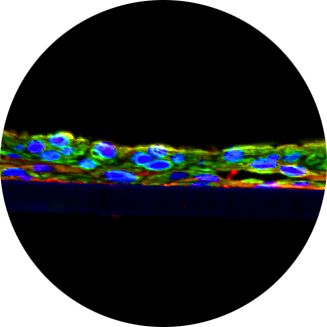
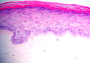
The Psoriasis tissue model is cultured using normal human epidermal keratinocytes and psoriatic fibroblasts harvested from psoriatic skin tissue specimens. The cells are cultured on specially prepared tissue culture inserts using serum free medium to form a multilayered, highly differentiated tissue. The reconstructed psoriasis tissues adopt a psoriatic phenotype as evidenced by increased basal cell proliferation, expression of psoriasis-associated biomarkers, and elevated cytokine release. The model provides researchers with a useful, in vitro means to assess the preclinical safety and efficacy of lead therapeutic compounds. For more information on the Psoriasis model, click here.
The Method
Evaluation of Psoriasis Drug Formulations Using a Human Tissue Model of Psoriasis
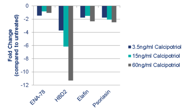
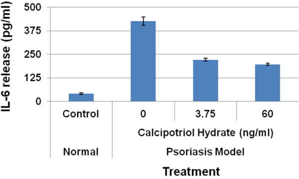
Mattek’s in vitro Psoriasis tissues were treated topically with 50µl of increasing concentrations of Calipotriol for 96 hours. Following treatment, tissues were processed for total mRNA isolation according to Mattek’s RNA Isolation Protocol. Analysis of psoriasis specific markers showed significant and dose-dependent reductions in Human beta-defensin 2 (HBD2), Elafin (PI3) and Psoriasin (S100A7) gene expression levels by qPCR. Additionally, analysis of cell culture supernatants showed a significant and dose-dependent reduction in IL-6 levels following treatment.
 Effect of calipotriol on psoriatic biomarkers – Repeat exposure (once daily) for 4 days
 Calcipotriol treatment: IL-6 release from Psoriasis tissue model
Effects of IL-17 on Chemokines and Cytokines that Mediate Chronic Inflammation in Psoriasis
As increasing evidence implicates the TH17 pathway as playing an important role in the pathophysiology of Psoriasis, the effects of IL-17 were investigated on Mattek’s Psoriasis tissue model in a tightly controlled in vitro setting. Exogenous IL-17 induced the secretion of chemokines and cytokines that mediate chronic inflammation in Psoriasis including CCL20 (MIP-3α, Th17 and Dendritic Cell chemoattractant), IL-8 (Neutrophil chemoattractant) and IL-6 (Th17 Cell survival). These results demonstrate that IL-17 exacerbates inflammation in an in vitro human cell-based model of Psoriasis.
Request a Quote
Thank you for requesting information about Mattek products! A representative will contact you shortly.
**If you would like to place an order for Mattek products, please contact Customer Service**


