Assessment of Alveolar Toxicity and Fibrotic Potential In Vitro using the Mattek EpiAlveolar™ Model
- TR Number: 845
- Authors: CS Roper, JE Baily, HJ Simpson, and JL Vinall
- Materials Tested: TGF-β, bleomycin, silica (50 nm)
- Institution: Mattek Corporation, Charles River Laboratories, Unilever
Pulmonary fibrosis (PF) is a debilitating, typically fatal, condition caused by a variety of factors including environmental or occupational exposures, drugs, radiation, and genetic predisposition. No current in vitro (immortalized cell lines), ex vivo, in silico or in vivo models of PF fully recapitulate all salient features of the human disease.
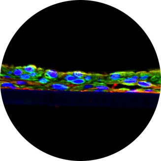
In this study, a novel in vitro organotypic 3D airway model from primary human cells (EpiAlveolar™ from Mattek Corporation) with macrophages was examined to evaluate alveolar toxicity and the capacity of the model as a method to assess fibrotic potential in vitro. Previously, this model has been shown to behave in a manner reflecting the parent tissue and under some experimental conditions can develop aspects of PF seen in the lower respiratory tract[1].
The study design was based on the protocol provided by Mattek[2]. EpiAlveolar™ tissues were challenged to develop aspects of PF, by exposure to known PF causing agents; bleomycin (12, 0.12 or 0.0012 µg/mL) or TGF-β1 (10, 5 or 1 µg/mL) in the culture media, or to repeated aerosol applications of silica (50 nm particle size, ca10 or 1 µg/cm2), for up to a 14 day period. Tissue viability was assessed by transepithelial electrical resistance (TEER) and lactate dehydrogenase (LDH) release every second day. Tissue samples and spent culture media were collected throughout the time course for analysis by pathology, immunohistochemistry (IHC) and ELISA to further characterise the healthy model and to identify aspects of PF developing over time.

