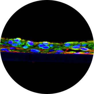
Organotypic in vitro human airway models can recapitulate aspects of pulmonary fibrosis
- TR Number: 844
- Keywords: Pulmonary fibrosis, EpiAlveolar, EpiAirwayFT, Cytokeratin 19, vimentin, Type I Collagen (COL1A1), Fibronectin (Fn), Tumor necrosis factor alpha (TNF-α), TEER, MMP-2, Type III Collagen, E-cadherin, Type I Collagen, α-smooth muscle actin (α-SMA), pro-fibrotic phenotype
Pulmonary fibrosis (PF) is a debilitating, typically fatal condition that may be caused by a variety of factors, including occupational and environmental exposures, drugs such as amiodarone and bleomycin, radiation exposure and genetic predisposition. However, in 20-30% of cases the cause is unknown (i.e. idiopathic pulmonary fibrosis, IPF). Currently approved IPF drugs (pirfenidone, nintedanib) have only had limited efficacy and lung transplantation remains the best treatment option for IPF patients.
Despite intense research, many of the molecular mechanisms involved in the initiation and progression of IPF remain unknown. Current IPF research relies on animal models and ex vivo lung tissues, which are expensive and are not always predictive of clinical trial results. In vitro models produced from immortalized cells also do not adequately replicate IPF.
The goal of the current work is to develop in vitro organotypic, 3D airway models from primary human cells which can be used to study PF.
Reference Application
- Absorption
- Antimicrobial
- Buccal delivery
- Genotoxicity
- Mucosal delivery
- Reproducibility - skin tissue models
- UV protection
- Corneal Drug Delivery
- UV damage
- transporters
- Regulatory Approval
- Review Article
- LOAEL
- Drug delivery
- Infectious disease research
- Buccal drug delivery
- Pharmacotoxicology
- Nasal absorption
- Nanotechnology
- Aging
- Respiratory Disease
- Bacterial infection
- Pollution
- Mildness Testing
- Barrier Disruption
- UVB
- Irritation
- Infection
- Mucosal irritation
- Psoriasis
- Vaginal irritation
- Respiratory immunotoxicity
- Human-on-a-chip
- Atopic Dermatitis
- DNA Damage
- Colitis
- Drug ADME
- Hair Growth
- scalp health
- Permeation
- Infections
- Cosmetics
- Metabolism
- Mucous
- Respiratory infection
- Viral Infection
- Melanogenesis
- Antiviral
- Gastrointestinal Disease
- Hazard assessment
- Biomedical Devices
- artificial saliva
- Skin irritation
- Toxicity
- Cytokine analysis
- Immulogical research
- Nanoparticle toxicology/penetration
- Respiratory toxicology
- MMPs
- Intestinal Permeation
- Bacterial colonization
- Translational toxicology
- Dry skin
- drug skin compatibility
- bacterial adherence
- Allergenicity
- Antioxidants
- Drug absorption
- Immunologicaal research
- Toxicology
- Skin cancer
- Photoaging
- Electrolyzed Water
- Transbuccal drug delivery
- XtraMild skin mildness testing
- Skin moisturization
- Protein Expression
- PBPK Modeling
- Immunological Research
- Apoptosis
- Intestinal toxicity
- Microbicides
- Nanotoxicology
- Skin corrosion
- Skin Sensitization
- Fibrosis
- Oral Pathology
- bacterial vaginosis
- ADME
- Gastrointestinal Inflammation
- anti-wrinkling
- Immunogenicity
- Basic dermal research
- Respiratory research
- Immunotoxicity
- Ocular irritation
- Penetration
- Medical Devices
- Pulmonary Fibrosis
- Oral infection
- vaginal microbiome
- Microphysiological system
- Gastrointestinal Irritation
- Nicotine pouch products
- Inflammation
- Asthma
- Skin corrosion Absorption
- Mucosal
- Oral candidiasis
- Skin lightening
- Organ-on-a-Chip
- Oxidative Stress
- Oral inflammation
- Collagen Remodeling
- Hyperpigmentation
- Barrier Function
- intranasal drug delivery
- Microbial infection
- COPD
- Smoking
- Respiratory toxicity
- Oral irritation
- Skin
- Oral Disease Research
- Skin Damage
- Ocular toxicology
- Drug Screening
- Skin de-pigmentation
- Gastrointestinal Toxicity
- permeation enhancement
- Wound healing
- Smoke
- Tobacco
- Inhalation Toxicology
- Oral mucositis
- Smoker
- Space Research
- Skin Barrier
- Validation
- Nephrotoxicity
- Cancer Research
- Skin Brightening
- excipients
- Phototoxicity
- Research
- Drug development
- Nanoparticles
- Skin pigmentation
- Gingivitis
- Dry Eye
- Drug Metabolism
- Biofilm
- Hepatotoxicity
- Personalized Medicine
- Food Additives
- Sub-categorization
- UV radiation
- Basic cutaneous research
- Epithelial restitution
- Microbial
- Pigmentation
- Transbuccal permeation/penetration
- Micronucleus Assay
- Skin aging
- Intestinal barrier
- Liver Toxicity
- Consumer products
- Biocompatibility
- Anti-aging
- Basic DC research
- Eye irritation
- Microbicide
- Pigmentation studies
- Oral mucosa
- microbiome
- Skin disease
- SARS-CoV-2
- Skin Toxicity
- Cytotoxicity
- reproducibility
- Skin hydration
- UV
- Genetic toxicology
- Microbicide testing
- Radiation
- Tumor invasion
- Probiotic
- Intestinal infection
- Skin re-epithelization
- Crohn's Disease
- Inflammatory response
- respiratory irritation
- UV toxicity
- Basic respiratory research
- Genomics
- STD infection
- Reproducibility - eye (ocular) tissue model
- UV light
- Irritation>Eye Irritation OECD TG 492
- Skin differentiation
- Barrier repair
- Inflamed Bowel Disease
- Visible Light
- pesticides
Reference Product
Ready to advance your science?
Our team is ready to provide a cost-free consultation to determine how we can help you reach your research and testing goals. Contact our team of experts today.

